


Research on Light Therapy and Parkinson’s Disease
Parkinson’s disease is a progressive neurological condition affecting movement, cognition, and quality of life. Early research into photobiomodulation is showing promising signals, exploring how light therapy may support brain function through its effects on mitochondrial health, blood flow, and inflammation.
Parkinson’s disease is a progressive neurological condition affecting movement, cognition, and quality of life. Early research into photobiomodulation is showing promising signals, exploring how light therapy may support brain function through its effects on mitochondrial health, blood flow, and inflammation.

Signs and Symptoms of Parkinson’s Disease
Symptoms of Parkinson’s disease include (Mayo Clinic, n.d.):
- Tremors
- Slowed movement, also called bradykinesia
- Rigid muscles
- Poor posture and balance
- Loss of automatic movements (ex. swinging arms while walking)
- Speech changes (slurring, sounding monotone)
- Writing changes
Biomarkers of Parkinson’s Disease
Neuron Loss in Parkinson’s
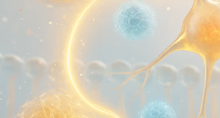
The most common biomarker of Parkinson’s disease is the ongoing death of dopaminergic neurons in the substantia nigra, which drives many of the symptoms (Surmeier, 2018).

Lewy Body Protein Aggregates
Fibrillar aggregates of Lewy Body proteins are found in many people with Parkinson’s disease and often contribute to the progression of the disease (Wakabayashi et al., 2007).

Detecting Alpha-Synuclein Clumps
A 2023 study of spinal fluid from over 1,000 people detected alpha-synuclein clumps, identifying Parkinson’s in 87.7% of cases and flagging those at risk (Siderowf et al., 2023).
What Causes Symptoms of Parkinson’s?
Oxidative Stress
An imbalance between the production of harmful reactive oxygen species) and the body’s ability to neutralize them with antioxidants, - plays a significant role in the degeneration of dopaminergic neurons leading to Parkinson’s disease (Meiser, Weindl & Hiller, 2013).
Mitochondrial Dysfunction
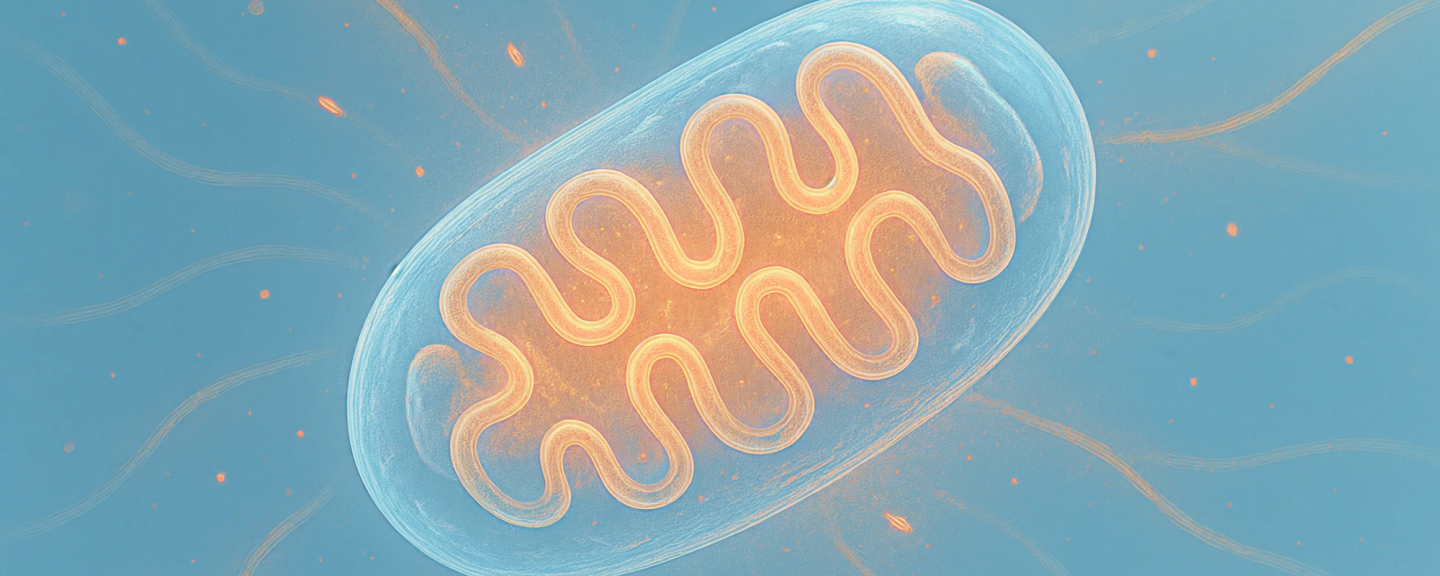
This dysfunction is highly implicated with Parkinson's disease. A reduction in adenosine triphosphate (ATP) production can lead to oxidative damage that harms lipids, proteins, and DNA and can further promote mitochondrial dysfunction (Pajares et al., 2020).
Scientific Insights - PBM’s Applications for Parkinson’s Disease
Clinical Benefits of PBM in Parkinson’s
Using near infrared light pulsed at 40 Hz showed improvements in mobility, cognition, dynamic balance and fine motor skill of people with Parkinson’s disease (Libert et al., 2021). Further, these results were sustained after both one and five years of use, suggesting that PBM may maintain improvements in the face of a degenerative disease.
See the studyPBM and Neurotransmitter Enzyme Activity
PBM has been shown to enhance the activity of enzymes like tyrosine hydroxylase, which play a key role in neurotransmitter synthesis, thereby supporting improved dopaminergic function in the brain (Moro et al., 2016).
See the studyPBM and Neurochemical Improvements in Rats
A 2023 study using rat models showed that PBM improved levels of dopamine, serotonin and norepinephrine that are implicated in the progression of Parkinson’s disease (Mohammed et al., 2023). Furthermore, rats treated with PBM also performed better on behavioral tests measuring locomotor activity.
See the study
FAQs
What results can be expected and when?
Types of results and their timelines vary depending on the individuals. However, a combination of research and anecdotal evidence from providers suggests some expected results to be the following: improved language capabilities, improvements in emotional regulation, decreased repetitive behaviours, and improved social skills/engagement. While some clients may see results very quickly, others may take a few months.
What is mitochondrial dysfunction?
Mitochondrial dysfunction is a term that encapsulates dysfunction in various operations of the mitochondria. With mitochondria being the main energy producers of the cell, dysfunction can lead to reduced ATP (energy) production, increased oxidative stress, and difficulty maintaining healthy cell function. When mitochondria can’t keep up with the body’s energy demands or fail to remove damaged components, it can affect brain function, muscle strength, immune response, and overall vitality. Mitochondrial dysfunction is linked to many conditions, including fatigue, neurodegeneration, and metabolic disorders (Zong et al., 2024).
How many PBM sessions will I need?
Benefits can often be noticed within a few sessions, especially related to sleep and mental clarity. For cognitive improvements, consistent use over several weeks or months is typically recommended. Neurodegeneration is often referred to as a progressive condition with genetic components; incorporating light therapy as a regular lifestyle habit can support overall brain health and give your brain the best chance to stay at its most performing level over time.
Is PBM safe?
Yes. PBM is FDA-cleared for various applications and has a strong safety profile. It’s non-invasive, painless, and has no known serious side effects when used as directed.
Learn More About PBM & Brain Health
 Blog
BlogMitochondrial Dysfunction: Causes, Symptoms, and How Light Can Help Restore Energy
Explore how PBM addresses mitochondrial dysfunction — restoring cellular energy, reducing oxidative stress, and supporting brain health

 Newsletters
The Most Overlooked Tool in Brain Injury Recovery and Biohacking
Discover why light therapy is becoming a key tool in brain injury recovery and biohacking—supporting energy, focus, and long-term resilience.

 Blog
BlogHow Brain Vital Signs and Photobiomodulation Are Shaping the Future of Brain Health
tPBM can rapidly improve cognitive processing, with cases showing up to 70 ms gains in healthy users, dramatic recovery post-concussion, and long-term benefits in mild impairment. Even advanced dementia patients show measurable brain activation changes.

References
Liebert, A., Bicknell, B., Laakso, E.-L., Heller, G., Jalilitabaei, P., Tilley, S., Mitrofanis, J., & Kiat, H. (2021, July 2). Improvements in clinical signs of Parkinson’s disease using photobiomodulation: A prospective proof-of-concept study. BMC Neurology, 21, Article 256. https://doi.org/10.1186/s12883-021-02248-y
Meiser, J., Weindl, D., & Hiller, K. (2013, May 17). Complexity of dopamine metabolism. Cell Communication and Signaling, 11(1), Article 34. https://doi.org/10.1186/1478-811X-11-34
Mohammed, H. S., Hosny, E. N., Sawie, H. G., & Khadrawy, Y. A. (2023, December). Transcranial photobiomodulation ameliorates midbrain and striatum neurochemical impairments and behavioral deficits in reserpine-induced parkinsonism in rats. Photochemical & Photobiological Sciences, 22(12), 2891–2904. https://doi.org/10.1007/s43630-023-00497-z
Moro, C., El Massri, N., Darlot, F., Torres, N., Chabrol, C., Agay, D., Auboiroux, V., Johnstone, D. M., Stone, J., Mitrofanis, J., & Benabid, A.-L. (2016). Effects of a higher dose of near-infrared light on clinical signs and neuroprotection in a monkey model of Parkinson’s disease. Brain Research, 1648 (Part A), 19–26. https://doi.org/10.1016/j.brainres.2016.07.005P
ajares, M., Rojo, A. I., Manda, G., Boscá, L., & Cuadrado, A. (2020). Inflammation in Parkinson’s Disease: Mechanisms and Therapeutic Implications. Cells, 9(7), 1687. https://doi.org/10.3390/cells9071687
Siderowf, A., Concha-Marambio, L., Lafontant, D.-E., Farris, C. M., Ma, Y., Urenia, P. A., Nguyen, H., Alcalay, R. N., Chahine, L. M., Foroud, T., Galasko, D., Kieburtz, K., Merchant, K., Mollenhauer, B., Poston, K. L., Seibyl, J., Simuni, T., Tanner, C. M., Weintraub, D., Videnovic, A., Choi, S. H., Kurth, R., Caspell-Garcia, C., Coffey, C. S., Frasier, M., Oliveira, L. M. A., Hutten, S. J., Sherer, T., Marek, K., & Soto, C.; Parkinson’s Progression Markers Initiative. (2023). Assessment of heterogeneity among participants in the Parkinson’s Progression Markers Initiative cohort using α-synuclein seed amplification: A cross-sectional study. The Lancet Neurology, 22(5), 407–417. https://doi.org/10.1016/S1474-4422(23)00109-6
Surmeier, D. J. (2018, August 14). Determinants of dopaminergic neuron loss in Parkinson’s disease. FEBS Journal, 285(19), 3657–3668. https://doi.org/10.1111/febs.14607
Wakabayashi, K., Tanji, K., Mori, F., & Takahashi, H. (2007). The Lewy body in Parkinson’s disease: Molecules implicated in the formation and degradation of α-synuclein aggregates. Neuropathology, 27(5), 494–506. https://doi.org/10.1111/j.1440-1789.2007.00803.x